
Cross-Sectional Atlas of the Rhesus Monkey Head: with 0.024-mm pixel size color images
- Publisher : Springer
- Illustrations : 250 col illus
Our customers have not yet submitted a review for this title - click here to be the first to write a review
Description:
The Visible Monkey is the first trial to obtain high-quality and real-color sectioned images of a rhesus monkey's whole body (intervals, 0.05 mm (head) and 0.5 mm (body except head); pixel size, 0.024 mm X 0.024 mm; color depth, 48 bits color).
This color atlas sets a new standard in rhesus monkey neuroanatomy by presenting around 400 ultrathin sectioned images of the head, including the brain, and whole body of the rhesus monkey. The image enabled observations of detailed anatomical structures, thanks to high-resolution and real-color sectioned images of the monkey unlike the stained sections and magnetic resonance images (MRI). Furthermore, a new reference system employed for this purpose is clearly explained for the readers, and structures are fully annotated in the horizontal, coronal, and sagittal planes.
Recent advances in 3 Tesla MRI and tractography from MRI have considerably enhanced imaging of the monkey brain, thereby impacting on both neuroscience research and clinical practice. Moreover, the information gained from initiatives involving photography of thin slices of cross-sectional images includes enriched knowledge of neuroanatomy and thereby facilitated the interpretation of such ultra-high-field resolution images. These exquisite images contained within this atlas will be invaluable in providing both researchers and clinicians with important new insights.
You may also like...

The Natural History of Crime: Case studies in death and the clues nature leaves
Wiltshire, P.
Price £22.00

Evolutionary Thinking Across Disciplines: Problems and Perspectives in
du Crest, A.; Valkovic, M.; Ariew, A.; Desmond, H.; Huneman, P.; Reydon, T.A.C. (Eds)
Price £119.99
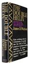
The Double Helix: A Personal Account of the Discovery of the Structure of DNA
Watson, J.D.
Price £10.00
La Nature: Revue des Sciences et de leurs Applications aux Arts et à
Tissandier, Gaston (Ed.)
Price £65.00

The Advent of PhyloCode: The Continuing Evolution of Biological Nomenclature
Laurin, M.
Price £89.99
